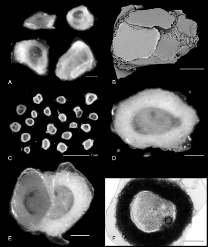
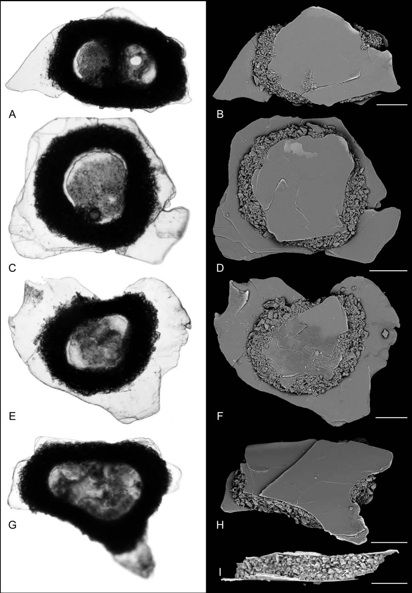
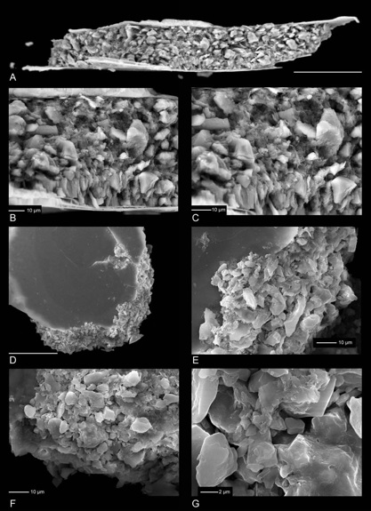
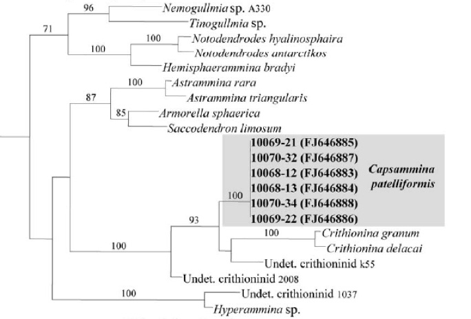
Capsammina patelliformis Gooday, Aranda da Silva, Koho, Lecroq and Pearse, 2010
Class Polythalamea
Order Allogromiida
Family Allogromida incertae sedis
Genus Capsammina Gooday, Aranda da Silva, Koho, Lecroq and Pearse, 2010
Species Capsammina patelliformis Gooday, Aranda da Silva, Koho, Lecroq and Pearse, 2010